In Which Organ Does the Vicious Cycle of Obesity Take Place?
I got a question on X: “In which organ does the vicious cycle of obesity actually take place?”
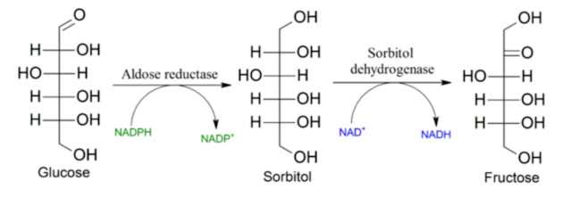
Well, I have to admit, those thoughts came so quickly that I haven’t really reflected on it much yet. The polyol pathway is active in the liver, but I’m not aware of its activity in adipose tissue. Fat synthesis takes place both in the liver and in adipose tissue. So for now, let’s stick with the hypothesis that it’s the liver. We actually have a nice study on a mouse model where they tested the knockout of fructokinase (KHK, the enzyme that activates fructose) both globally in the organism and separately in the digestive tract or in the liver. The results are very interesting.

Turning off fructose processing in the digestive tract prevents fructose from entering the body fairly well; it also seems to suppress the craving for sweets and reduces the consumption of sweetened water. But it does not prevent metabolic syndrome or weight gain in any way. The result is fatty liver and obesity. There is always enough fructose in the body to manifest itself and cause metabolic syndrome.

In contrast, turning off fructose processing in the liver (KHKFl/FlXCre-Alb) is quite a different story. Take a look at the figure above. Fructose intake is not disrupted at all; total calorie intake even increased by the amount of fructose. But since it can’t be processed into anything useful, it passes through the body into the urine and is excreted.
What happened to body weight? We don’t have a graph, but the result is clear enough. While the control group gained about 10 grams, the group lacking fructokinase in the liver gained nothing at all. The liver and the whole body have the same weight as the control group. The liver is healthy and fat-free. There was no increase in insulin or leptin levels. Everything is normal even on a glucose-fructose water in the diet. The differences between the groups are not only in the liver but throughout the entire body. We can see this in the adipose tissue—the size of fat cells is normal, not enlarged. Cellular senescence was not examined, but I dare say they are not senescent because, as the authors point out, there are no crown-like structures around them, no signs of damaged or dead fat cells.
This fits quite nicely into the model I have already presented here. Daily food intake is regulated by excess carbohydrates and proteins not processed by the body, which are converted into glucose and stored as liver glycogen. This provides the body with information about its energy balance.

The liver can synthesize new fats, combine them into triglycerides, and export them as VLDL particles, which ensure their storage in adipose tissue. In this way, it can distribute calories between consumption and storage. We can see this activity in full swing in this study. The main switching signal is the level of hepatic hydrogen peroxide (H₂O₂), which triggers fat synthesis and export. The main stimulant for H₂O₂ production is fructose created via the polyol pathway, as I described in the previous post. In cases of digestive tract problems or overload, dietary fructose itself can serve as this signal.
This signal can be further enhanced by the presence of the aldehyde HNE, which is a product of omega-6 fatty acid peroxidation. So I wondered, could the enhancement of the effect in the digestive tract enhance the desire for sweets, since the absence of KHK reduces the preference for sweetened water in mice?

Fructose, through AMP molecules, activates the AMPD2/XO/uric acid pathway. In addition to the fact that the activation of the KHK-C enzyme suppresses antioxidant protection, the enzyme XO releases H₂O₂ during its activity. That is enough to activate fat production in the liver and to redirect incoming calories from immediate use toward long-term fat storage. And that is the foundation of fat accumulation not only in the liver but throughout the entire organism. Simply flip this caloric switch back toward consumption, and fat no longer needs to be stored—fat cells are not overloaded, and the entire organism becomes much healthier. Substances like acetate/vinegar can help with this.
The problem, moreover, is that redirecting calories toward storage leads to increased food intake and a higher need for rapid fat storage in adipose tissue. Without available acetate, this leads to adipose tissue damage. If overeating occurred with natural food—as nature intended—part of the food would reach the large intestine and return to the body as acetate, a product of the gut microbiome. Acetate has the ability to activate the deacetylase SIRT1 precisely at the time when it is necessary to increase the number of active fat cells—when stem fat cells need to differentiate. Without this mechanism, adipose tissue damage and cellular senescence occur.

References:
Counteracting Roles of AMP Deaminase and AMP Kinase in the Development of Fatty Liver
Role of AMPD2 in impaired glucose tolerance induced by high fructose diet


Comments
Post a Comment