How exactly do omega-6 oils affect metabolism? Is vinegar/acetate really the culprit?
Let me start with some reasoning because there’s still a lot of confusion surrounding the metabolism of seed-based vegetable oils, which primarily contain the omega-6 fatty acid linoleic acid (LA). Studies show very mixed results, which is why irreconcilable camps of supporters and opponents of seed oils have formed among different dietary preference groups. You already know my stance, but I always try to remain as unbiased as possible first. This post is meant as a hard blow to researchers because everything might be completely different from what we’ve thought so far. The reason is that our researchers often forget to measure some basic metabolic parameters, which then secretly skew the results in one direction or another far more than the observed variables. But more on that later.
A while back, I explained that when linoleic acid is burned in the mitochondria, there’s an increased consumption of NADPH molecules because the enzyme 2,4-dienoyl-CoA reductase (DECR) is activated. I need to revisit this issue because it’s not the only pathway that leads to higher NADPH consumption. The metabolism of linoleic acid differs slightly between mice and humans, and this has consequences.
To process fatty acids, three basic enzymes break down long chains into shorter two-carbon segments: VLCAD, LCAD, and MCAD. LCAD handles chains with 12 to 18 carbons, while VLCAD deals with longer ones. But human mitochondria, unlike mouse mitochondria, lack LCAD, so mice can burn linoleic acid in their mitochondria without much trouble, while humans struggle a bit. There are two ways to deal with this issue.
The first option is to activate peroxisomes via PPARα and burn linoleic acid there. This can be triggered, for example, when LA is accompanied by other exotic, rarely occurring fats — like the omega-3 α-linolenic (ALA) acid from flaxseeds or even better EPA and DHA from fish oil. This activation kicks peroxisomes into gear and produces acetate, which is often used to synthesize new fats. As Brad Marshall correctly identified, this whole process acts as a filter that safely processes these exotic fats.
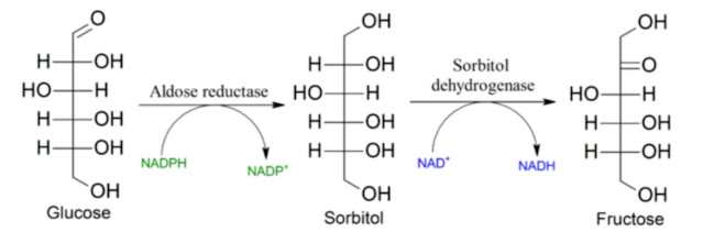
The second option is to elongate and desaturate linoleic acid to make it more compatible with VLCAD, allowing it to be burned in mitochondria. We know this is possible from studies on diseases linked to peroxisomal genetic disorders. LA isn’t processed directly, so its levels rise significantly, but its elongated byproducts remain at normal or even lower levels. So, elongation and desaturation help. Elongation consumes NADPH, while desaturation consumes NADH.
What does this tell us? NADPH consumption isn’t just increased due to DECR processing LA — elongation also requires NADPH. Additionally, this process needs malonyl-CoA, meaning de novo lipogenesis (DNL) must also be turned on. So, if we shut down DNL (e.g., by activating AMPK), we reduce LA burning in mitochondria and the harmful effects of higher NADPH consumption in them — but at the cost of elevated LA levels, which should theoretically allow for safe peroxisomal LA burning. Interestingly, turning off DNL only redirects LA. This could be useful and might relate to the effects of a ketogenic diet, which strongly suppresses DNL. Yet, from experience, we know that LA increases ketone production (which happens in mitochondria), so maybe the shutdown isn’t 100% effective. Or does LA independently trigger DNL? I don’t know.
Brad Marshall also pointed out that linoleic acid allows glucose fermentation to continue without lactate buildup — simply because polyunsaturated fats (PUFAs) can be desaturated, freeing up NAD+ to sustain glycolysis for ATP energy. That’s fine, but it reinforces the problem: LA not only reduces NADPH but also lowers the insulin resistance we want to preserve (which is protective) while increasing the insulin resistance we don’t want (stem cell senescence in fat tissue). There are multiple types of insulin resistance, after all. But is PUFA desaturation the dominant factor here? Maybe not for us. We’ll see.
Now, let’s take a closer look at a study where rats were fed an extremely high-fat diet rich in seed oils (Dyets #112245: ~0–1% myristate, ~5% palmitate, ~2% stearate, ~12% oleate, ~80% linoleate; 60% fat, 15% protein, 25% carbs). It took me a while to track down the fat composition, but I finally did. Unfortunately, I couldn’t confirm whether the diet contained fructose. This study suggests things might work differently. The key player? Acetate — and a neural gut-brain-pancreas connection via the vagus nerve. Acetate is rarely measured in studies, even though it’s a major metabolic actor.
 |
| Acetate turnover in the body depending on the caloric intake of rats. |
The processing of LA begins in the small and large intestines, where bacteria break it down. As Brad Marshall hinted, oxygen is used for desatureation — PUFAs facilitate glucose fermentation even in lactate-rich environments, which would otherwise stall the process. So, with enough PUFAs in the gut, glucose fermentation happens much more easily, producing high acetate levels. Other studies show that omega-3 fats (with more double bonds) are the best substrate for acetate production, while small amounts of omega-6 tend to suppress gut bacteria. But in high amounts, omega-6 seems to directly activate acetate-producing bacteria.
 |
| Acetate boosts insulin secretion (insulin AUC) during glucose stimulation and is a product of gut bacteria — antibiotics completely block acetate production, but acetate infusion restores it. |
Interestingly, these researchers seem to dislike acetate, describing it as a harmful byproduct. Strange, considering how much we’re told to nurture gut bacteria, especially those producing short-chain fatty acids (like Akkermansia muciniphila).
 |
| Is acetate really the culprit? |
 |
| Gut bacteria as perpetrators of acetate turnover after 4 weeks of high-fat diet with vegetable oils, GF germ-free, CONV-D implanted with standard microbiome. |
The study found something fascinating: acetate reaches the brain, where it activates neural pathways that increase insulin secretion (GSIS) in response to food. Acetate appears during overeating and helps store excess calories. Blood sugar stays more stable (no spikes), but at the cost of higher insulin levels. Whether this is better is debatable. If it increases protective insulin resistance while reducing fat stem cell senescence, it might be beneficial. Overeating is an easily solvable problem — just slow down food intake and improve diet quality. Acetate actually suppresses hunger, so the overeating in this study was likely due to the artificial diet (probably containing sugar, which activates KHK and DNL). Removing fructose from the diet and creating a harder form of the diet should solve this. I wouldn't see that as a major problem. Note that if the rats didn't overeat, everything was the same as on the standard diet, with the same bacteria.

 |
| Acetate caused no problems when rats were pair-fed. |
Key quote:
„Pair-feeding with isocaloric portions of chow or HFD produced no change in acetate turnover or GSIS...“
But the researchers concluded that acetate is bad — causing hunger, overeating, and obesity. This contradicts previous studies showing the opposite. But it doesn’t matter because we already know that hunger is ultimately regulated by liver glycogen, likely via the brain.
 |
| When acetate was infused into the stomach, it caused overeating (or was it fructose?). But vagotomy (cutting the vagus nerve) erased acetate’s effects. |
If overeating is prevented, even this absurdly high-PUFA diet (60% calories from 80% linoleic acid oil) is protected by acetate, likely produced via glucose fermentation in the presence of unsaturated fats. The ability to desaturate PUFAs is key here — it allows acetate to lower blood glucose during overeating and facilitate fat storage. The study itself admits: if overeating is avoided, this high-PUFA diet is essentially equivalent to standard rodent chow. That’s huge, right? We don't have to cut our vagus nerve for this, anyway, the first overeating was caused by a sudden change in diet to a very fatty one. Just don’t overeat — not even short-term. Slowing calorie intake is crucial.
How many studies on PUFA metabolism have you seen? A lot? And how many measured acetate turnover and overeating? None? Exactly. This is the first study to actually examine PUFA metabolism while tracking acetate. Without this, all other studies are useless — just speculation and errors. As a layperson and engineer, I have to say it bluntly: this is bad science. We learn almost nothing applicable from them. This study is groundbreaking.
Addition:
Unlike rodents, the effect of acetate on brain stimulation and insulin secretion is opposite in humans, acetate reduces insulin levels in obese people.

References:
Acetate mediates a microbiome–brain–β-cell axis to promote metabolic syndrome
Mitochondrial long chain fatty acid beta-oxidation in man and mouse
The short-chain fatty acid acetate reduces appetite via a central homeostatic mechanism


Comments
Post a Comment