What do lactate and fish oil have in common? They suppress obesity! Why?
Lactate is a product of glucose fermentation, usually thought to cause muscle fatigue and problems, but probably not at all. Fish oil is known for containing a large amount of omega-3 fats, we will mainly be interested in the longest omega-3, i.e. DHA. What could these two substances have in common, that they suppress the development of obesity. Attention! Let's distinguish suppressing of obesity development from treatment, i.e. let's distinguish prevention of obesity from elimination of already existing obesity, these are two completely different disciplines!
Let's first look at the study with fish oil, i.e. with DHA. Here we have a mouse model study that includes four types of high fat diet. Control low-fat (Con, 10% fat), high-fat diet with lard (HFD, 60% fat), high-fat diet with lard and safflower oil (SO, 30%+30%), high-fat diet with lard and safflower oil and fish oil (SF , 30%+15%+15%), and finally a fatty diet with lard and fish oil (FO, 30%+30%).
 |
| Fish oil (SF, FO) acts with lifelong use as a prevention against omega-6 fats that shut down the malate-aspartate shuttle. |

 |
| High expression of IDH2 signals insufficient function, insufficient restoration of glutathione, lack of NADPH. |
Obesity problems appeared only in the groups without fish oil. Both groups of mice fed a high-fat fish oil diet showed no signs of obesity. Why? I think you can see for yourself with the graphs. It is the enzyme isocitrate dehydrogenase 2 (IDH2), i.e. the mitochondrial enzyme for the recovery of NADPH, which is part of the TCA cycle. While the amount of the enzyme is practically the same in all groups with a high-fat diet, gene expression, i.e. the requirement for protein production, is many times higher in the two obese groups. From this I conclude that in these two groups more IDH2 protein/enzyme would be needed, but the capacity to synthesize it is not available. Therefore, NADPH is not produced enough for proper metabolism, not enough to restore the level of the main cellular antioxidant glutathione, this causes an increased production of the H2O2 signal and the cell reacts with a defense mechanism, i.e. excessive fat production.
We see that not all fats behave the same, certain fat composition can be obesogenic. By slightly changing the composition of fats, an obesogenic diet can be adjusted to a normal, non-obesity-causing diet. What the study does not tell us is that an obesogenic diet will make more or less permanent changes in adipose tissue, and later adjustments to the composition of the diet will no longer have the desired effect. The correct composition of fats must be administered from an early age. An obesogenic diet is one that produces too much H2O2 when burned in the mitochondria, a signal of excess fuel or lack of oxygen. As we already know, adaptation to a low-oxygen environment, pseudohypoxia, is triggered, and HIF1A is activated in some fat cells.
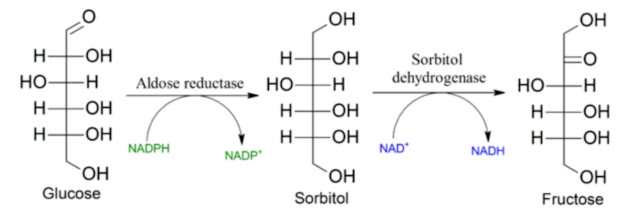
What makes DHA so essential? It activates the burning of fats in peroxisomes, supports their formation and reduces the load on mitochondria. The result of beta oxidation of fats are NADH and acetyl-CoA molecules, but lactate, malate and acetate emerge from peroxisomes. This is due to the impermeability of the membranes to NADH. Auxiliary shuttles, lactate or malate-aspartate, are used to export energy (electrons) from peroxisomes. The malate-aspartate shuttle can restore NADH in the cytosol, or it can be extended into the mitochondrion to restore the NADH level there. For us, the restoration of the NADH level in the cytosol is more important because, as we will see later, it regulates the supply of glucose.
And now we move on to lactate. A study has recently been published that surprisingly finds that lifelong lactate supplementation is protective against obesity. It turns out that lactate produced by muscles during exercise can prevent negative changes in the metabolism of mice on a high-fat diet. So how is it? No positive result occurs in mice fed a normal low-fat diet, but lactate shows a protective effect when fed a high-fat diet. It certainly cannot repair an already damaged metabolism, but it works as a prevention. Similar to active movement and exercise. Interestingly. So let's look at the results in more detail, let's try to find the mechanism.
 |
| Lactate supplemented throughout the lifetime counteracts obesity caused by certain fats (omega-6 linoleic acid) |
Controlling processes that run one after the other (in a serie) is not a simple matter. Mr. Ford once invented a conveyor belt system to avoid accumulation of unfinished products at intermediate operations. Nature cannot use any conveyor belt, therefore it has to regulate each process separately and even over one or two processes. Such a system is difficult to tune, and any violation of one regulation will cause changes in all regulatory loops.
Let's go back to the diagram of glycolysis. This time we will completely ignore the regulation via insulin and AKT, because in my opinion this is a higher regulation that only reflects the damaged regulation of the lower levels.

If we are to look for the heart of the problem with the fat diet of mice, this is how I see it. We know that for some reason that we are not interested in right now, the activity of de novo lipogenesis (DNL) is higher, so the ACC enzyme is more active, and the malonyl-CoA created by it prevents long fatty acids from entering the mitochondria for being burned a little more. It simply burns less fat than corresponds to the composition of the diet. So there must have been a little less activation of AMPK, and that's because of a little higher level of fructose-1,6-bisphosphate (Fru1,6BP). Fru1,6BP turns out to be essential for detecting sufficient glucose in the cell and for regulating the entry of glucose into the cell, or its phosphorylation to glucose-6-phosphate (Glc6P).
So the question is, how is the level of Fru1,6BP regulated? It is the molecule that is in the middle of the process. Its level is determined by the activity of input and output enzymes. The input enzymes are controlled by the level of Fru1,6BP in a negative feedback loop, so the level is stabilized. The uptake of molecules to farther processes is determined by the activity of the GAPDH enzyme, its function seems to be key. Thus, a higher level of Fru1,6BP could be a manifestation of low GAPDH activity.
 |
| A high-fat obesogenic diet suppresses GAPDH activity more than NADH levels (probably by high H2O2 production). |
As with all similar enzymes, the main regulator of GAPDH activity will be the level of the produced cofactor, i.e. NADH. A low level of NADH will increase GAPDH activity and allow more glucose to be replenished. A high level of NADH will do the opposite. If only glucose is burned, the level of NADH is lower, GAPDH activity is higher, Fru1,6BP is massively taken up for further processing, and glucose phosphorylation proceeds normally at the input.
When burning glucose with fat, the level of NADH will increase and the entry of glucose will be "choked" via GAPDH and a higher level of Fru1,6BP so that everything is well managed. The criterion for proper functioning of the regulation is usually low mitochondrial H2O2 production during oxidative phosphorylation. If we overload the mitochondria, they will let us know by increasing the production of H2O2, and the cell must respond to this. An appropriate response is to increase the oxygen supply, another appropriate response is to activate mitochondrial uncoupling or reduce the amount of fuel. We already know that if the cell does not react in time, the metabolism switches to fermentation. HIF1A is activated. This triggers the activation of the immune system and the activation of NOX2 enzymes, which in turn produce H2O2 and try to shut down the cell by apoptosis. Often, instead, there is only a decrease in GAPDH enzyme activity and an increase in Fru1,6BP levels, a decrease in AMPK, an increase in ACC, and an increase in de novo lipogenesis and a reduction in fat burning. Unless HIF1A is deactivated by acetylation, this state is permanent for the fat cell.
I can't help it, the main regulator of glucose entry into the cell is the cytosolic level of NADH. If the mitochondrial level of NADH ceases to be copied to the cytosolic via the malate-aspartate shuttle, regulation of glucose uptake ceases to function. Processes that reduce the level of NADH must not exceed certain limits, i.e. not even the consumption of NADPH. It is used to restore the main cellular antioxidant glutathione (GSH). There are fats that use up more GSH than other fats. There are even fats that directly consume NADPH (omega-6) during beta oxidation. With a high-fat diet, NADPH production consumes NADH or limits mitochondrial NADH production. A higher consumption of NADPH will therefore result in a decrease in the level of NADH. This reduction is transferred to the cytosol, where NADH is also reduced, presumably to replenish glucose from which NADPH is produced via the PPP pathway. But the capacity to produce NADPH from glucose is not enough to burn fats, especially polyunsaturated fats with double bonds in even positions. The cell wants more NADPH, it lets in more glucose, but the PPP pathway to NADPH is already saturated, so glucose reaches the mitochondrion, replenishes the NADH level, but the antioxidant system is overloaded and the H2O2 level increases. The cell's response is either a short-term restriction of GAPDH with activation of DNL, or a long-term restriction of the main pyruvate entry into mitochondria, the PDHC enzyme, through activation of HIF1A (in fat cells). Both methods break the link between the mitochondrial and cytosolic levels of NADH by lowering the levels of both aspartate and malate, thus breaking the aspartate-malate shuttle. In order to restore the levels of aspartate and malate, it is necessary to create oxaloacetate from pyruvate with the help of the mitochondrial enzyme PC, which will supplement the aspartate-malate shuttle with the necessary molecules. However, the entry of pyruvate into the mitochondrion is blocked (PDK activated by HIF1) or pyruvate is not formed at all (blockage of GAPDH by H2O2). In case of HIF1 activation, lactate formation is started by LDH, this is rescue fermentation metabolism, pseudohypoxia.
 |
| Extracellular lactate restores the malate-aspartate shuttle and cytosolic NADH levels. Thus, it prevents flooding of mitochondria with excess fuel, excess pyruvate, limits GAPDH activity and reduces H2O2 production. |
So how can lactate help us avoid this state?
Lactate available from the metabolism of other cells or supplemented externally slows down or reverses the action of LDH towards the formation of NADH and pyruvate. It will therefore help to keep the aspartate-malate shuttle running and thus maintain the level of cytosolic NADH even when burning problematic fats that consume a lot of NADPH. This is important because the NADH regulatory ability of GAPDH will be preserved and the aspartate-malate shuttle will not be disconnected by the lack of pyruvate and the subsequent activation of HIF1A in some fat cells, which will then trigger permanent obesity.
Let's go back to DHA and fish oil. If we know that DHA activates and multiplies peroxisomes, and if we know that the output of beta oxidation of fats in them is malate and lactate, the mechanism is the same as when supplementing with external lactate. It just needs pyruvate or oxaloacetate from the mitochondria. It works as prevention, but DHA cannot deactivate the already activated HIF1A signal in fat cells. Conversely, in a strongly oxidizing environment (NOX2), it can be degraded into toxic auto-oxidation products. Neither fish oil nor DHA is suitable as a dietary supplement for pre-existing metabolic problems. HIF1A and NOX2 must be turned off first!
And one more addition.
I also slightly overlooked the part in the study with lactate that first fed the mice a fatty diet for 8 weeks and then tried to make them lose weight with intensive exercise or intensive exercise supplemented with lactate injection. And they succeeded. This part of the study represents a model of treatment, not just prevention. This confused me a bit, I point out above that treatment and prevention are two different disciplines and so far the only substance/substances that really cure obesity are short chain fatty acids (SCFA), most often acetate. I know of no other such substance. So from this point of view, we should scrutiny every successful weight loss study to see if acetate could somehow have done it. Unfortunately, no obesity and weight loss study ever measures either acetate levels or fluxes. Never! Is it intentional?
So the question is, could the above studies be successful in preventing obesity precisely because of the effects of acetate? Could the acetate flows and levels have changed in any way? And the answer is: Yes. Acetate could have caused it.
In the first study with fish oil, it could be increased hepatic production of acetate during peroxisome activation, additionally associated with lactate production, the effect of which I will mention later.
As such, lactate should make the situation worse. If we have mice on a diet consisting mainly of sugar (approx. 10% cal) and lard (60% cal), then the intestinal bacteria produce enough endotoxin LPS. Fats have the unpleasant property of getting these endotoxins into the internal circulation, into the body. These endotoxins then bind to TLR4 receptors and cause inflammation. We are mainly interested in inflammation in adipose tissue. In this blog, I mostly write about so-called pseudohypoxia, i.e. the activation of HIF1A =HIF-1α and the activation of the NOX2 enzyme, which is ultimately the same as inflammation. The pseudohypoxic cell calls the immune system and the result is inflammation, but it is not activated by TLR4 receptors. Whether TLR4 or HIF1A activation is the cause cannot be easily distinguished because they trigger each other and vice versa. And lactate should increase the activity of TLR4, so it should make the situation worse. Why does it improve it?
Here I have to ask myself, can't acetate also play a major role in lactate study? It can! Lactate activates the MCT1 membrane transporters, increasing their number in the cell membrane. These transporters are very good at transporting not only lactate, but also acetate. I don't have data on this, but in vivo, that is, in a living model, the level of acetate in the cell can change significantly. This can affect HIF1A acetylation and turn off pseudohypoxia and inflammation. I don't know how lactate could do this other way than through acetate. Increasing the level of NADH or pyruvate alone is certainly not enough to turn off HIF1A and NOX2.
 |
| Extracellular lactate (red) will increase number of MCT1 transporters. They can let not only more lactate, but also more acetate into the cell, which can turn the fermentation metabolism back to the oxidative one where possible. However, this requires the presence of extracellular acetate |
Acetate may only function in the presence of extracellular lactate. This would mean that it is the hypoxic cells after the end of hypoxia that will be the biggest recipients of acetate. For acetate to be effective as a therapy, lactate levels might also need to be increased. This can be done relatively easily with fructose, for example, honey or fruits. Or maybe through exercise, or by red light therapy. Or slow nasal breathing.

It will be necessary to further investigate the mechanisms, it would be good if the researchers take the effects of acetate in consideration and always evaluated when studying obesity whether the positive effects could be caused by acetate or another SCFA. Then maybe we could get to a real understanding of obesity.
References:
Docosahexaenoic acid mediates peroxisomal elongation, a prerequisite for peroxisome division
Kinetic modeling of glucose central metabolism in hepatocytes and hepatoma cells
Fructose-1,6-bisphosphate and aldolase mediate glucose sensing by AMPK
Mechanism of GAPDH Redox Signaling by H2O2 Activation of a Two−Cysteine Switch


Comments
Post a Comment