Do AMPK phosphorylation and ATP level control vascular calcification?
The strengthening and hardening of soft tissues by calcification is a very interesting phenomenon. The same process takes place in the strengthening of bones, but is usually undesirable in the case of soft tissues. Actually, it is not calcium as such at all, but calcium phosphate (hydroxyapatite, HA, Ca10(PO4)6(OH)2), which strengthens soft tissues. It is the very same material that is missing from the bones in osteoporosis, while it builds up and damages the soft tissues. The driving force of calcification is therefore not calcium at all, but phosphorus, or inorganic phosphate (Pi). And to make it not so simple, it depends on its form. Inorganic phosphate (Pi) accelerates calcification, inorganic pyrophosphate (PPi) protects against calcification. The difference or ratio between the levels of these two forms of inorganic phosphorus is stabilized and balanced by a group of enzymes called alkaline phosphatases (ALP/TNAP). These break down the pyrophosphate into two phosphate molecules that promote calcification. Extracellular PPi is formed mainly from ATP molecules that leave the cell and release PPi when AMP is formed. A sufficient level of protective PPi can only be maintained if ALP/TNAP activity is low. If ALP/TNAP is activated, calcification will occur. Of course, all these processes are strictly regulated, and the PPi molecule therefore serves as a signaling molecule. What does it actually signal? I would say enough ATP. And that brings us to the basic problem. Could the calcification be caused by a lack of ATP, insufficient phosphorylation of AMPK and ACC1, which would direct fuel processing towards fat storage and cause a lack of ATP for other processes?
 |
| Tissue non-specific alkaline phosphatase (TNAP/ALP) breaks down pyrophosphate into inorganic phosphate, nucleoside pyrophosphatase (NPP1) separates two phosphates from ATP and creates AMP, which activates AMPK phosphorylation and ATP generation. It is therefore a closed cycle that is influenced by molecules that block AMPK phosphorylation. |
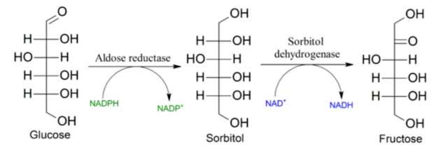
I have drawn attention here several times to the basic problem of metabolism, the phosphorylation of AMPK and ACC1 enzymes. If we have enough food and ACC1 is not phosphorylated, we will get fat. Food will be stored as fat. The enzyme that phosphorylates ACC1 is the AMPK enzyme. So we need to phosphorylate AMPK to not get fat, to burn fuel and make cellular energy ATP. Thus, the entire regulatory loop appears to be phosphor-dependent, comprising ATP production, extracellular PPi, breakdown of PPi to Pi, and AMPK phosphorylation. This cycle is controlled by other substances that prevent phosphorylation. One of them is the sugar detection system through fructose-1,6-bisphosphate and aldolase enzyme. The excess of carbohydrates blocks the phosphorylation of AMPK, turns on the mechanisms of protection against excess glucose in the blood, i.e. the conversion of glucose into fat and its storage. This in itself is fine, but you also need to turn off this process in time.
Another such molecule is aldosterone. There will probably be more such molecules. What is interesting is that if we prevent the phosphorylation of AMPK, there will not only be an increase in the production of new fats, but also the so-called autophagy, cleaning in the cell, will be turned off. Degradation of old non-functional enzymes and materials will be prevented. This also causes calcification of soft tissues. We can show it in a study with aldosterone. It prevents the phosphorylation of AMPK in the same way as carbohydrates do.
 |
| Aldosterone (Aldo) blocks the phosphorylation of AMPK, so blocks autophagy, i.e. the controlled removal of non-functional proteins and structures. This creates conditions for calcification of blood vessels. |
This regulation of calcification works by aldosterone suppressing AMPK phosphorylation. An increased level of inorganic phosphate Pi should normally increase the phosphorylation of AMPK, this would raise the production of ATP and the protective PPi, but this does not occur. Calcification will occur, i.e. the formation of hydroxyapatite. If we remove the AMPK phosphorylation blockers, ATP production increases and calcification is prevented.
 |
| Chronic elevation of aldosterone (DOCA) modulates calcification only with increased phosphate uptake (HPD). No effect without phosphate. The activity of the enzyme alkaline phosphatase (ALP/TNAP) follows the calcification curve. |
 |
| Aldosterone causes calcification only when phosphate levels are elevated. |
 |
| Calcification is increased by the addition of phosphate (Pi), further increased by the addition of aldosterone and phosphate (Pi+Aldo), blocking the aldosterone receptor will reduce calcification (Pi+Aldo+Spiro) because AMPK phosphorylation is not inhibited, suppressing AMPK will reverse the effect of blocking the receptor and trigger calcification (+ Compound C). |
We can show that calcification is triggered in mice by increasing phosphate intake to about three times the normal intake. This underlying calcification can be further modulated by supplying aldosterone and blocking aldosterone receptors. As this modulation is mediated by AMPK phosphorylation, a similar increase in and suppression of calcification can be achieved by modulating AMPK phosphorylation. That brings us to the effects of vinegar/sodium acetate. It can also modulate AMPK phosphorylation. We don't know exactly how it does it, but we do know that the activation of acetate to acetyl-CoA releases pyrophosphate from ATP, and thus the product is phosphate for phosphorylation.
| Before oxidation to CO2 and H2O |
| After oxidation to CO2 and H2O. |
I have discussed the effects of sodium acetate on metabolism recovery many times. But what effects does it have on soft tissue calcification?
There is a study investigating the effects of high blood acetate concentration in connection with dialysis (artificial kidney). The normal level of acetate in the blood is about 0.1 mmol/L. In the case of supplementation of approx. 200 mg/kg, the level can reach up to 1 mmol/l. Here, however, they tested levels of about 20 mmol/L, because that is the concentration of solutions used historically in dialysis using acetate solutions. Today probably completely different solutions are used for dialysis, the results with acetate are interesting for us. At such high concentrations, soft tissue calcification occurs. Why? Because there will be a significant increase in circulating phosphorus. The mechanism of acetate activation is to blame.
 |
| Note the increase in pyrophosphate (PPi), especially about 100x with acetate infusion while fasting (starved). Pyrophosphate normally has a protective effect, with an acetate infusion of up to 20 mmol/L it already has a negative effect on calcification. |
Note that pyrophosphate is released. Several hundred such events, when pyrophosphate is released, take place in the cell. In the case of acetate activation, its positive effects on AMPK and ACC1 phosphorylation can be explained in this way. But a very high level drives phosphorus out of the cell, into the extracellular space, where it triggers calcification. It is therefore advisable not to exaggerate the concentration of acetate during supplementation. The processing of approx. 20 g of sodium acetate (trihydrate) takes about an hour and produces a concentration similar to the consumption of alcohol. There, however, the concentration of acetate is roughly constant throughout the alcohol processing period. It is likely that excessive use of acetate can have similar effects on the liver as excessive alcohol consumption, fattening and hardening the liver.
 |
| Sodium acetate will increase resting energy expenditure exactly by the values given by burning the acetate. It does not appear to limit the burning of other fuels, i.e. carbohydrates or fat. |
 |
| Time course of blood acetate concentration in humans after ingestion of approximately 20 g of sodium acetate (trihydrate). |
At low enough concentrations, vinegar/acetate can counteract soft tissue calcification by activating AMPK phosphorylation. But high concentrations can cause the exact opposite. A high level of extracellular phosphate directly causes the formation of hydroxyapatite. In order to maintain the protective effect of pyrophosphate, the activity of the enzyme alkaline phosphatase (ALP/TNAP) must be suppressed. It is the activity of ALP/TNAP in different tissues that determines whether they will be hard (bones) or soft (vessels). The activity of the ALP/TNAP enzyme depends, among other things, on the concentration of H2O2, oxidative stress activates ALP/TNAP, thus increasing the level of extracellular phosphate and contributing to a considerable extent to the calcification of soft tissues. This is how the effect of oxalate on blood vessels can be explained.
References:
Fructose-1,6-bisphosphate and aldolase mediate glucose sensing by AMPK
The medical and metabolic consequences of administration of sodium acetate


Comments
Post a Comment