Hydrogen Sulfide – Regulator of Gluconeogenesis, Vascular Health, Liver, Muscles, Adipose Tissue, etc.
We continue with the effects of hydrogen sulfide on enzyme activity. We have already seen that H₂S affects the S-sulfhydration of sirtuins, i.e., the activity of deacetylases that change the activity of many enzymes by preventing their “decoration” with an acetyl group. In the case of sirtuin modification, for example, this facilitates the “decoration” of some enzymes with ubiquitin, which leads to their removal. Or it increases their activity after the removal of acetyl “decoration,” if that decoration interferes with the enzyme’s function. Even at this level, things are already quite complicated. But that’s not all. S-sulfhydration of certain sites can occur on many proteins, not just on sirtuins! Research shows that H₂S also sulfhydrates the enzyme pyruvate carboxylase (PC) and also the key metabolic switch, the enzyme AMPK. That would explain further phenomena observed both in vitro (in the test tube) and in vivo (in whole organisms) related to changes in hydrogen sulfide levels. S-sulfhydration of cysteine—i.e., the modification of a built-in amino acid in an enzyme in such a way that the -SH group is changed to an -SSH group, thereby preventing “decoration” at that site—is thus a very powerful enzyme modification. It is striking how little this is generally known, because it fundamentally changes our understanding.

 |
| The influence of H₂S depends on many factors; in different tissues it can be very different—for example, in muscles it depends on whether we are dealing with slow oxidative fibers or fast glycolytic fibers. |
Allow me to quote from one interesting study:
„S-sulfhydration modification involves in the regulation of glucose metabolism, cardiovascular system, inflammation, oxidative stress, mitochondrial biosynthesis, apoptosis, endoplasmic reticulum (ER) stress, and DNA damage repair. The regulation of H₂S on AMPK activation has been reported in heart, H9c2 cells, and colonic muscle cells.”
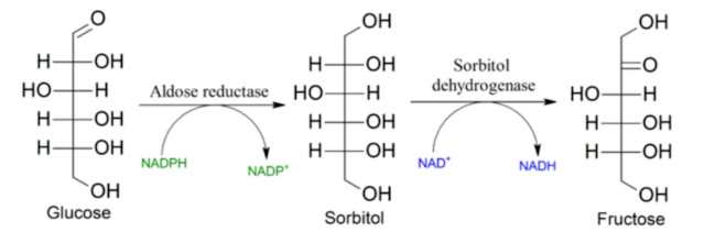
In this post we will look at how hydrogen sulfide affects glucose metabolism in muscles and how it affects gluconeogenesis in the liver. I have already hinted at my hypothesis that daily food intake is controlled by the amount—or perhaps rather the changes in the amount—of liver glycogen so that after a day’s exertion we have enough glycogen for the period of rest and sleep. That is the daily task for us as well as for animals: we must have enough energy for sleep. And precisely the influence on gluconeogenesis is therefore related to the amount of food we eat. If hepatic hydrogen sulfide S-sulfhydrates pyruvate carboxylase (PC), its activity increases, especially when food is abundant, with high levels of acetyl-CoA. Higher PC activity caused by hydrogen sulfide thus reduces our need to eat more.
 |
| Activation of pyruvate carboxylase (PC cDNA) activates gluconeogenesis, the formation of glucose from lactate or amino acids. Suppression of hydrogen sulfide production (CSE-KO), as well as suppression of PC (PC-siRNA), turns off gluconeogenesis. |
Another enzyme strongly influenced by S-sulfhydration is AMPK. Recall that this is a kinase, i.e., an enzyme that phosphorylates other enzymes, which directly phosphorylates the enzyme ACC. This is the main fuel switch. Phosphorylation of ACC decreases its activity and, in times of plenty, prevents the creation of new fats and instead promotes greater burning, i.e., energy expenditure rather than storage. Low AMPK activity in times of plenty thus leads to storing and conserving fat, preparing for winter. Hydrogen sulfide, by activating AMPK, signals to cells that they don’t need to conserve and store energy, but can afford to waste it.
 |
| Hydrogen sulfide activates AMPK phosphorylation and glucose metabolism. |
As we already know, our modern diet forces us to overeat; restriction of sulfur amino acids should therefore, through activation of hydrogen sulfide production from homocysteine via the enzyme CSE, raise hydrogen sulfide levels in various organs and induce changes that are different in each organ. In the liver, it leads to better glycogen storage and thus prevents overeating. In adipose tissue and in muscle it activates AMPK, which leads to energy being wasted as heat and to lower fat storage. Whether the same mechanism is also activated by overall calorie restriction of protein below 10%, I don’t know, but it is quite likely.
 |
| High levels of glucose (HG) or free fatty acids (FFA) deactivate hydrogen sulfide production by the enzyme CSE. |
Another organ regulated by hydrogen sulfide is our vasculature, specifically its ability for vasodilation, i.e., regulation of blood flow and pressure. This process is controlled by nitric oxide (NO) and the enzyme eNOS, which produces NO from arginine. S-sulfhydration of eNOS can significantly increase NO production and thereby improve vascular function.
 |
| Cysteine, via the enzyme CSE, increases hydrogen sulfide levels, improves endothelial function, and enhances nitric oxide production. However, regular supplementation with cysteine probably suppresses the activity of the enzyme CSE and leads to chronic hydrogen sulfide deficiency. |
Since the main enzyme producing up to 90% of hydrogen sulfide is CSE, it is necessary to examine how to maintain its activity under various conditions.
 |
| Hydrogen sulfide produced by CSE reduces fat and cholesterol synthesis in the liver. Switching off CSE (CSE-KO), combined with choline deficiency (CD), shuts down H₂S production and thus activates fatty liver disease. |
Animal models clearly show that high blood glucose, as well as high free fatty acid levels, lead to a loss of CSE function and a decrease in hydrogen sulfide levels. The cause is mitochondrial overload with fuel—whether carbohydrates or fats. Do you now see why cells must initiate insulin resistance? Whether it’s common physiological insulin resistance or the ultimate result of chronic overload, i.e., cellular senescence. The reason is that overload causes increased oxidative stress, which causes oxidation of membrane phospholipids and release of 4-hydroxy-2-nonenal (4HNE). That attaches to enzymes and causes loss of hydrogen sulfide production.
 |
| 4-hydroxy-2-nonenal (4HNE) suppresses the function of detoxifying/aldehyde-removing enzymes (ALDH2), antioxidant defense enzymes (G6PD), and deacetylating enzymes (SIRT2). |
Can reducing intake of omega-6 linoleic acid help? If throughout life we have not restricted intake of seed-derived vegetable oils such as sunflower oil, then our adipose tissue has stored very large reserves of linoleic acid. If one of these molecules oxidizes to the aldehyde 4HNE, it is released and immediately is replaced by a new molecule—we have plenty. The only option is to reduce oxidative stress, i.e., repair oxidative metabolism, and simultaneously activate clearance via the enzyme ALDH2. It seems that hydrogen sulfide is exactly what we need for this. S-sulfhydration is so protective that it repairs acetylation and even nitrosylation of enzymes.
 |
| High protein consumption, i.e. exposure to high levels of cysteine, likely suppresses H₂S formation in the long-term between meals. |
In my previous post, I showed the protective effect of activating CSE and hydrogen sulfide. If CSE can be activated by reducing intake of cysteine and methionine, then probably the simplest approach is to reduce intake of all amino acids, i.e., to lower dietary protein. But how to prevent muscle breakdown? Won’t we be deficient in protein? It is probably necessary at the same time to reduce the use of amino acids for energy, i.e., for gluconeogenesis. The simplest compensation for the lack of dietary protein is glucose. It might seem that carbohydrates are best, but they usually come with hidden proteins. So what provides energy without protein? Sugar. There are indeed enthusiasts of sugar diets who successfully use them, but as I have analyzed here before, this is not a method for the general obese population. These people are dependent on fat burning for survival. Fructose causes severe hunger in them by blocking fat utilization. Therefore, I propose using pure glucose as a dietary supplement.
An interesting observation is that activation of CSE, i.e., increasing hydrogen sulfide levels, not only activates oxidative phosphorylation—better burning of fats and glucose—but also activates PC and gluconeogenesis. This can be good for fat burning because it activates the malate-aspartate shuttle for NADH/NAD⁺ exchange and citrate production. But in combination with glucose? Fortunately, PC activity is strongly dependent on acetyl-CoA concentration. A low-fat diet is associated with the creation of new fats precisely from citrate, which is converted by the enzyme ACLY into acetyl-CoA. So if everything is functioning properly, the acetyl-CoA level will not be high enough to activate gluconeogenesis by H₂S.
 |
| A striking analogy between the effects of sulfur amino acid (SAA) restriction and the effects of hydrogen sulfide (H₂S). |
References:
H2S-induced S-sulfhydration of pyruvate carboxylase contributes to gluconeogenesis in liver cells


I've been loving the articles.
ReplyDeleteH2S is interesting because I saw research about a year ago saying to much H2S in the gut leads to overeating by suppression of GLP-1. (actually robs cells of energy and the lack of E stops production)
https://www.nature.com/articles/s42255-024-01068-x
https://www.gastrojournal.org/article/S0016-5085(98)70311-7/fulltext#:~:text=A%20simple%20approach%20to%20reducing,release%20of%20H2S.
Fiber, is another option, with all it's other benifits
https://pubmed.ncbi.nlm.nih.gov/26203099/
Thank you, studies on mouse diets could be misleading, hardly applicable to humans. Many parameters should be controlled, but no one cares to measure or control them.
DeleteI would say my N=1 has been successful.
ReplyDeleteTook about two weeks at 525mg a day bismuth subsalicylate for it to kick in. Presumable, long term damage to gut needs time to heal. But then after that, my experience is that hunger response to meals is much stronger. I'll quit half way through a meal. It's a very different experience to feel full now. You are not supposed to take it long term, so not sure where to go with it.
Randomly found this human trial of sulfide reduction.
https://translational-medicine.biomedcentral.com/articles/10.1186/s12967-023-04833-w#:~:text=Dietary%20sulfur%20amino%20acid%20restriction%20(SAAR)%20has%20been%20investigated%20in,%5B12%2C13%2C14%2C