The Rate of Glucose Phosphorylation as a Cause of Insulin Resistance?
Once again, insulin resistance. I have already discussed this phenomenon several times here — a phenomenon many scientists consider the cause of civilization diseases, especially obesity and type 2 diabetes. We have shown that insulin resistance (IR) is an overly general term that does not capture the various mechanisms of its formation — that IR has its own causes and is therefore a reaction to some condition, an evolutionarily developed defense mechanism, generally beneficial and thus cannot be the root cause.
Since then, I have discussed several other potential root causes of IR on this blog. In the previous post, I was intrigued by the issue of glucose phosphorylation. Although it seems like a simple process, it hides many complexities. I’ll show you a paper in which the author examines this problem in detail. It demonstrates the relationship between glucose transporters GLUT4 and hexokinase HK2 and insulin. In the resting state, with low insulin levels, glucose transporters GLUT1 are the limiting factor. Upon activation by insulin, GLUT4 transporters move to the cell membrane, and glucose transport ceases to be the limiting factor. This is where the rate of glucose phosphorylation by enzymes HK1 and especially HK2 becomes the new limiting factor. The author, however, is unaware of the polyol pathway — of the cascade of events triggered by activation of the KHK/AMPD2 enzyme, of the uric acid production, and of a drop in ATP generation. He also doesn’t know that deactivation of KHK synthesis prevents metabolic issues associated with sugar and high-fat diets. Yet, it is precisely the limiting activity of HK2 — and thus the low rate of glucose phosphorylation — that is likely the main activator of the polyol pathway when insulin levels are high.
When you start exploring how HK2 works, you find that glucose phosphorylation occurs through the conversion of ATP to ADP. A cofactor, G6P, must also be present — but it’s also the product of the reaction and inhibits it when it accumulates. This seems simple until you discover that HK1/HK2 can, but don’t have to, be bound to the outer mitochondrial membrane via VDAC1/ANT — meaning they may have direct access to ATP produced by oxidative phosphorylation in mitochondria. It therefore appears that the relocation of HK2 between the membrane and cytoplasm can significantly alter its properties, especially its limiting rate.
 |
| The localization of HK2 on the mitochondrial membrane ensures rapid glycolysis to pyruvate; localization in the cytosol leads to slower glycogen production. |
This is also supported by another study examining the effect of positive ions on HK2 activity. The authors found that neither calcium nor sodium ions affect HK2 activity, but potassium ions (K⁺) strongly activate the rate of phosphorylation — but only when HK2 is bound to the outer mitochondrial membrane. When in the cytosol, this effect disappears. It therefore seems that localization of hexokinase on the mitochondrial membrane is crucial for maintaining a sufficient phosphorylation rate under insulin-stimulated metabolism. This also has another effect — the process of glucose phosphorylation lowers the potential of the inner mitochondrial membrane and reduces ROS production. Glucose thus acts as an antioxidant, helping to prevent peroxidation of sensitive polyunsaturated fats. Furthermore, G6P is a substrate of the PPP pathway for NADPH recycling — the energy source for the glutathione antioxidant system. So, what regulates HK1/HK2 localization? It appears that a lack of fuel promotes binding to the membrane.
 |
| Calcium, magnesium, and sodium have no effect on the rate of glucose phosphorylation in the cytosol, but potassium removal stops glycolysis. |
In the fasting state, HK1/HK2 molecules are mostly bound to the mitochondrial membrane. Food intake increases glucose and insulin levels. Short-term insulin stimulation (around 30 minutes) strongly activates HK2, but as glycolysis products accumulate, HK2 detaches from the membrane. Further insulin stimulation (>4 hours) no longer increases phosphorylation speed — HK2 separates from the membrane, and the phosphorylation rate becomes limited.

HK2 localization likely also determines whether glucose is used to produce pyruvate or glycogen. Membrane-bound HK2 facilitates glycolysis and pyruvate formation, lowers the inner mitochondrial membrane potential, and thus reduces superoxide (O₂⁻) and hydrogen peroxide (H₂O₂) production.
Cytosolic HK2, on the other hand, favors glycogen synthesis. This relocation is mainly regulated by G6P levels — the product of hexokinase activity. After quickly phosphorylating glucose entering the cell under insulin stimulation, HK2 detaches from the membrane, loses access to the rapid ATP supply, and continues slower glucose phosphorylation for glycogen synthesis. An elegant system — but problems may arise when overloaded.
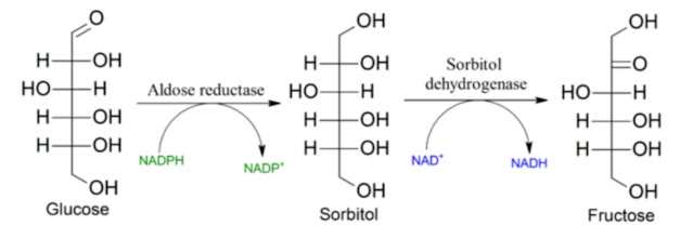
Let’s summarize the system’s properties. The polyol pathway functions as a safety mechanism against overload — a limiter. Like a boiler thermostat that shuts off fuel supply if the temperature exceeds safe limits, in our case it monitors cytosolic glucose levels. When these exceed the activation threshold for aldose reductase, glucose activates KHK/AMPD2, suppresses oxidative phosphorylation, and triggers glucose fermentation to lactate and fat storage. It limits fuel entry into mitochondria and favors cytosolic processing. This is a clear adaptation to an environment where cells are rapidly flooded with glucose they cannot process quickly enough. The fallback mechanism is always storage.
What about the interaction between hexokinase and protein metabolism? This is likely a complex relationship — but let’s simplify. Deamination of excess nonessential amino acids, especially glutamine and glutamate, releases ammonia, which changes cytosolic pH. Excess ammonia induces pseudohypoxia. Could hexokinase be involved? A cytosolic pH change from 7.4 to 6.0 immediately causes HK1 to relocate from the mitochondrial membrane into the cytosol. The need to process excess nonessential amino acids (e.g., converting them into fat for storage) thus leads to ammonia release, pH changes, HK relocation, and decreased glucose phosphorylation rate. Is polyol pathway activation merely a result of glucose overload — or also of amino acid metabolism? Possibly both. We know that limiting protein intake increases metabolic rate — and thus glycolysis speed.
 |
| Deamination of glutamine/glutamate in mitochondria releases ammonia and lowers cytosolic pH. |
But why do we overeat? I believe it’s related to amino acid availability — particularly essential amino acids (EAA), whose deficiency is life-threatening. In such cases, the energy-regulating feeding system becomes secondary, overridden by an emergency mechanism driving increased food intake to secure enough EAA. This can easily explain general problems of modern diets and matches what we observe in animal models.
The difference between healthy and unhealthy diets may lie only in structure, preparation, and absorption rate. For example, hard carbohydrate pellets are healthy for mice, but softened or finely ground ones are not, even though they have identical composition. The only difference is that they can be eaten and converted to glucose more quickly. Similarly, proteins can be healthy if they are fully utilized by the body. The same protein quantity but with a different amino acid composition can cause overeating if it triggers EAA deficiency signals (e.g., by flavoring with monosodium glutamate). Ultimately, the whole problem can be simplified as an issue of overeating. However, it’s not willpower-driven overeating — it’s caused by activation of protective emergency mechanisms evolved for times of food or protein scarcity, when the body defends itself from potential deficiency by promoting overeating. This in turn triggers other emergency processes linked to fat storage — and thus obesity.
 |
| Mice on soft food gain weight equally on both low-fat and high-fat diets. C-H/C-S = control hard/soft pellets; HF-H/HF-S = high-fat hard/soft pellets; W-H/W-S = obesogenic Western diet in hard/soft pellets. |
References:
Insulin, Muscle Glucose Uptake, and Hexokinase: Revisiting the Road Not Taken
Potassium ions promote hexokinase-II dependent glycolysis
Hexokinase-I directly binds to a charged membrane-buried glutamate of mitochondrial VDAC1 and VDAC2
Causes and Consequences of A Glutamine Induced Normoxic HIF1 Activity for the Tumor Metabolism


Comments
Post a Comment