Are High Blood Glucose Levels Harmful, Even Without Aldehydes?
The effort to lower blood glucose levels seems obvious, the reason is simple. High glucose levels cause oxidative stress and damage to body organs (eyes, blood vessels, kidneys, etc.). This leads many people to promote and apply low-carb and ketogenic diets.
But is it really glucose that causes it? After all, it is the most common fuel for obtaining energy so that cells can function at all. Isn't that strange? Moreover, the cell can easily defend itself against overload by means of insulin resistance. So why doesn't it work?
Is it really glucose that is harmful to the body?
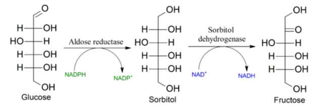
So let's take a closer look. If you are reading this blog, I think you already know where the core of the problem is. Yes, it is the activation of an enzyme that is supposed to serve only in emergency situations, otherwise it should be deactivated. It is the enzyme aldose reductase (AR, AKR1B1). It is present in the liver, vascular endothelium, cornea, etc. It is the first enzyme of the so-called polyol pathway, the product of which is fructose made from glucose.
AR should be inactive under normal conditions with normal glucose levels. But this is often not the case. It is also activated by oxidative stress. And this can be induced in many different ways. For example, by an excess of oxalates, by choline defitiency, or by deficiency of cis-vaccenic and palmitoleic acid, or by various toxins.
I think the mechanism of AR activation by oxidative stress is clear. First, the products of autooxidation of polyunsaturated linoleic acid are released from the membranes, especially 4-hydroxy-2-nonenal (HNE). This directly or as a conjugate with GSH (GS-HNE) activates AR and is converted by it into substances that cause inflammation. This pathway is part of the defense mechanisms, but it should not be activated permanently. In addition, fructose formation is also triggered, so the enzyme KHK and all the pathways associated with it are also activated.

In this way, a signal is activated in the long term to redistribute available fuels more for storage than for immediate consumption. We know the consequences. It is obesity, lack of cellular energy for maintenance and repair of cells, reduction of body temperature, etc.

Let's look at a 2019 study in which scientists investigated the effect of AR on the resilience of vascular endothelial cells. I have already discussed a similar issue here several times, we learned that acetate acts as a protector of vascular endothelial cells against senescence, etc.
How is this related to the AR enzyme and the activation of the polyol pathway?
I would say that the main pathway leads through fructose and the KHK enzyme, which turns off the SIRT1 deacetylase, although fructose is not mentioned even once in this study. It is a pity that these connections are not generally known. We clearly see here in the last column of the graph that the harmfulness of high glucose levels (HG) can be suppressed precisely by turning off the production of aldose reductase (AR, AKR1B1 siRNA). We also see that this is manifested by the activation/phosphorylation of the pAMPK/AMPK kinase, the basic cellular energy switch.
I will at least list the titles of the paragraphs with the results (HUVEC = human umbilical vein endothelial cells):
- AR inhibitor fidarestat prevents HG-induced HUVECs death and ROS formation
- AR inhibition prevents HG-induced apoptosis markers in HUVECs
- Fidarestat prevents HG-induced monocyte adhesion and endothelial dysfunction
- AR inhibitor fidarestat regulates Sirt1 expression in HUVECs
- Fidarestat regulates HG-induced AMPKα1 and mTOR activation
- AR ablation prevents HUVEC death and activates SIRT1 and AMPKα1
- AR regulates the expression of SIRT1 and phosphorylation of AMPKα1 and mTOR in heart and aorta tissues of STZ-induced diabetic mice

High glucose (HG) levels increase intracellular oxidative stress, that much is clearly visible. But is glucose really to blame? If we suppress the function of the AR enzyme with its chemical inhibitor (Fid), high glucose levels do not fundamentally matter. It is therefore purely a matter of the presence of substances produced by this pathway, the effect of sorbitol or fructose or both substances together.
The authors of the study tried various approaches, genetic or pharmaceutical blockade of AR (siAKR1B1, Fidarestat), cell tests and tests on mice. It always showed the same results. If the polyol pathway is not specifically deactivated, the result is oxidative stress (i.e. peroxidation of polyunsaturated fatty acids), suppression of SIRT1 deacetylase and suppression of AMPK activity (and an increase in AMPD2 and uric acid formation, which is not mentioned here).

At the center of this process is the SIRT1 enzyme, which also deacetylates enzymes associated with the production of NADPH molecules (G6PD, IDH2). NADPH molecules serve, among other things, to recycle antioxidant protection using glutathione molecules (GSH/GSSG). The SIRT1 activator (Resveratrol, Res) as well as the AR deactivator (Fidarestat, Fid) suppresses the effect of excess glucose, while the SIRT1 blocker (SIRT1 inh) reduces the viability of vascular endothelial cells.
Why does the suppression of the AR enzyme make the cell more resistant to excess glucose? This is because the AR processes unphosphorylated glucose. Once glucose is phosphorylated in the cell, it will be used by other pathways. I have already described the problem of the rate of glucose phosphorylation here.
High glucose levels reduce cell viability, but AR suppression immunizes cells so that high glucose levels do not bother them. This protection is given by the activation of the deacetylase SIRT1, the abundance of NADPH molecules, the preservation of antioxidant protection even under these conditions.
So what is the assumed mechanism that keeps repeating itself here? Do you see the loop, the positive feedback?
⬆️ glucose -> ⬆️ AR -> ⬆️ sorbitol ->
-> ⬆️ fruktose -> ⬆️ KHK -> ⬇️ SIRT1 ->
-> ⬇️ NADPH -> ⬇️ GSH/GSSG ->
-> ⬆️ oxidative stress -> ⬆️ PUFA peroxidation ->
-> ⬆️ HNE -> ⬆️ AR -> ⬆️ sorbitol etc.
From this diagram it is clear that suppressing AR breaks the loop.
We can continue, however:
⬆️ HNE -> ⬇️ ALDH2 -> ⬇️ AMPK ->
-> ⬆️ HNE -> ⬇️ ALDH2 etc.
So another loop, both loops are interconnected via HNE. ALDH2 activation removes HNE in a safe way and works very similarly to AR deactivation and it would be best to try to combine both ways. This should basically prevent many metabolic problems even in situations where there is too much linoleic acid in the membranes that causes this loop.
References:
Aldose reductase regulates hyperglycemia-induced HUVEC death via SIRT1/AMPK-α1/mTOR pathway


Comments
Post a Comment