What Is Lipofuscin and How To Avoid Its Production?
As the organism and its cells age, a substance called lipofuscin accumulates in the cell cytosol. It is a yellowish-brown metabolic waste (pigment) formed by oxidized fats and proteins. It is commonly called the “pigment of aging” because it gradually accumulates in cells (especially in neurons, heart muscle, liver and retina) and its amount naturally increases with age. It is not a homogeneous compound, it is probably a mixture of intertwined and interconnected chains that exhibit fluorescence. This fluorescence when stimulated by near-UV radiation is not always the same, it varies depending on the place of origin, composition and age from green to blue.
The literature tells us that it is actually unprocessed cellular waste. Normally, these substances would be removed by the formation of lysosomes, organelles where unusable substances are digested and broken down into reusable material or waste. Lipofuscin is the remnant of dysfunctional lysosomes, so it shows a state where the decomposition was not completed and substances were formed that cannot be decomposed under the given situation. So they remained encapsulated in lipid vesicles.
What affects the formation of lipofuscin? I will provide information here from a not too old study that summarizes the results of many studies on lipofuscin, especially from the perspective of the retina, which is very metabolically active and is very sensitive to the accumulation of lipofuscin. If you are interested in the details, read the entire study, it is quite long and detailed.
Quote
"Consistently, a decrease in lipofuscin accumulation can be achieved by upregulation of autophagy by inhibition of the mammalian target of rapamycin complex 1 (MTORC1), by rapamycin, or by pharmacological activation of the transcriptional control of transcription factor EB (TFEB), which stimulates the expression of autophagy proteins, lysosomal membrane proteins, and lysosomal hydrolases.
An increased lipofuscin accumulation is also a characteristic feature of many, but not all, diseases associated with increased oxidative stress, and it has been demonstrated in numerous experiments in vitro and in vivo that accumulation of lipofuscin can be accelerated by increased oxidative stress. For example, it has been shown that lipofuscin accumulates more rapidly in cultured cells or in animals exposed to increased oxygen tension, redox-active iron ions, or depleted of antioxidants.
Based on experimental findings on cultured rat cardiomyocytes, Brunk and colleagues proposed that autophagy and lysosomal degradation of organelles producing reactive oxygen species and containing redox-active iron ions, such as mitochondria, can increase oxidative stress by facilitating lipid peroxidation. Subsequently, the formation of oxidized and crosslinked biomolecules makes them no longer susceptible to lysosomal degradation. Iron chelators and antioxidants, such as vitamin E, can effectively inhibit lipofuscin accumulation by preventing lipid peroxidation."
End of quote

Lipofuscin accumulation can therefore be easily induced by oxidative stress or enzymatically, e.g. by activating the mTOR factor. On the other hand, lipofuscin accumulation can be suppressed by a substance called rapamycin, which blocks the mTOR signal. If we use the information from my recent post, we see that accelerated lipofuscin accumulation can be triggered by the aldehyde HNE. This blocks the AMPK kinase and thus activates the mTOR signal, which stops autophagy (removal of cellular waste) and blocks the work of lysosomes.
High-quality antioxidant protection, which reduces the peroxidation of linoleic acid to HNE, is therefore a basic condition for limiting lipofuscin accumulation in cells.
I can't help but think of the same model again. It is necessary to maintain a functional glutathione recycling system, sufficient vitamin E and sufficient retinoic acid, i.e. vitamin A.
What prevents this the most?
I'll repeat myself, it's still the same song. The presence of aldehydes and dysfunctional enzymes for their removal, specifically the presence of the linoleic acid peroxidation product HNE (4-hydroxy-2-nonenal) and the dysfunction of the ALDH2 enzyme, or in this case perhaps more of a problem with cytosolic ALDH1A1, which is an enzyme that activates vitamin A. Although it is not directly deactivated by the presence of HNE, it is dependent on deacetylation by the SIRT2 enzyme.


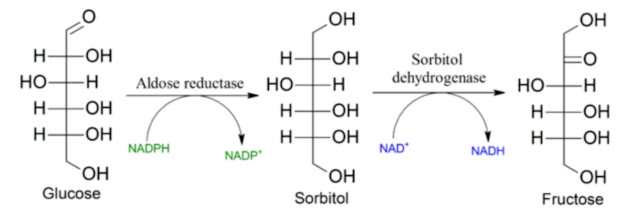
But SIRT2 is repressed by KHK activation, i.e. activation of AR by the aldehyde HNE (4HNE). Thus, the effect of HNE on ALDH1 (ALDH1A1, RALDH1) is indirect through the polyol pathway and aldose reductase (AR) activation.

In addition to the oxidative stress caused by activation of the polyol pathway by HNE, it also leads to a decrease in the conversion of retinal to retinoic acid (the active form of vitamin A). The active AR enzyme also directly removes retinal and further reduces the availability of active vitamin A. Activation of the polyol pathway and the KHK enzyme triggers oxidative stress by suppressing glutathione recycling due to the lack of NADPH molecules. This activates an environment favorable for the formation of lipofuscin.
It therefore seems that the peroxidation of linoleic acid to HNE plays a major role in the production of lipofuscin. Suppression of the AR enzyme (e.g. with glycine) ensures deacetylation of the enzymes and higher activation of the ALDH1 and ALDH2 enzymes, which should ensure greater availability of vitamin A, activate the AMPK enzyme and thus suppress the mTOR signal and restore autophagy, i.e. the processing of cellular waste by ensuring the proper functioning of lysosomes.
References:
NOTCH-induced aldehyde dehydrogenase 1A1 deacetylation promotes breast cancer stem cells


Comments
Post a Comment