How Can Linoleic Acid in Adipose Tissue Be Safely Replaced with Oleic Acid?
In one of the previous posts, we saw how the omega-6 vegetable fat stored in adipose tissue, specifically linoleic acid (LA), causes weight gain even with reduced food intake when blue light induces increased lipolysis. Eventually, it is probably depleted and replaced by oleic acid, a much more stable fat. At least, this is how I explain the rather incomprehensible recovery of metabolism at the end of the experiment. It did take somewhat longer than the approximately 20 days that might be expected based on the results of the study of LA kinetics in mice, but the change was induced solely by ambient light, not by zero fat intake.

The 1959 study of LA kinetics in mouse adipose tissue is interesting because it shows how, after just three weeks on a diet providing 30% of calories from vegetable oil, the fat composition of fat cells changes to become very similar to the composition of the oils being consumed. It is as if the composition of dietary fats were copied directly into the composition of adipose tissue. There are very few differences. After returning to a fat-free diet, the stored LA is replaced relatively quickly by oleic acid produced from starches through the process of de novo lipogenesis. The overall ratio of unsaturated to saturated fats remains almost constant, at approximately 70% / 30% in adipose tissue, whereas the ratio in the oils consumed is approximately 80% / 20%. But these are mice; in humans, the whole process probably proceeds much more slowly — it takes years rather than weeks.


Only corn oil (CO) is somewhat anomalous; it differs in some way from the other two oils. We can see this by comparing the data with a table of oil compositions. Metabolism typically replenishes only about 6% of oleic acid through de novo lipogenesis, which occurs with safflower oil (SO) and peanut oil (PO). But corn oil replenishes twice as much, approximately 13%, and more than 40% of the linoleic acid is probably converted into oleic acid before it is stored. This is therefore considerably more than with the other oils.
What could explain this? Why do some dietary oils trigger a faster transformation of LA into oleic acid?
This appears to be a very useful process. Could it be caused by greater activation of the PPARα factor? Some trace fat may activate it, possibly the omega-3 fatty acid α-linolenic acid (ALA), because it seems to disappear in a rather unusual way. Corn oil contains 1.1% ALA, but adipose tissue contains only half that amount, approximately 0.55%.

Let's return to the blue-light study discussed in the previous post. Although metabolism returned to normal at the end of the experiment, there were also side effects. For example, the liver was probably poisoned by the aldehyde 4-hydroxy-2-nonenal (HNE), a toxin derived from LA peroxidation. Although we observed this indirectly, we can infer with reasonable confidence what happened from the metabolic data. Last time, I did not show you the liver or kidney damage.

Although the liver weighs less, it shows clear signs of fat accumulation, while the kidneys show signs of degeneration. I think both are consequences of HNE aldehyde poisoning.
Could excessive release of fat from adipose tissue (and therefore weight loss) be dangerous?
I think that the rapid release of linoleic or arachidonic acid from fat cells may be dangerous to our organs, including the liver, kidneys, and heart, precisely because oxidative stress transforms these fatty acids into aldehydes. Enzyme poisoning by HNE molecules is no joke. We do not know whether the mice on the zero-fat diet suffered liver and kidney damage. The 1959 study does not tell us that.
So how can we safely eliminate LA from adipose tissue without damaging vital organs?
I have already outlined one method here. It involves replacing some of the long-chain fatty acids in the diet, and therefore in the blood, with short- or medium-chain fatty acids. But phospholipids are probably not the main place where linoleic acid needs to be replaced. Triglycerides, which form the body's main reservoir of LA, appear to be more important. In other words, the LA stored in adipose tissue.
So how can linoleic acid in triglycerides be safely replaced with oleic acid? And how can LA be released from adipose tissue?
In mice, researchers achieved this relatively quickly using a special, unnatural diet completely free of fat, based on albumin and starch. I don't know, but it reminds me of experiments with sugar-based diets. After all, even the effect of consuming fiber (fructooligosaccharides) is essentially the effect of slowly replenishing blood sugar, glucose. No long-chain fats, only de novo lipogenesis. But a zero-fat diet is probably neither tasty nor safe for our organs.
Another, probably more common method is fasting, which lowers insulin levels. The main issue will be how well the released LA can be processed, since it preferentially enters the liver and kidneys, where it is converted into acetyl-CoA, which is then used to build palmitic acid. This is subsequently elongated and finally desaturated into oleic acid. This is therefore a process of replacing vulnerable LA with safer oleic acid, so that it can be safely burned for energy or stored in adipose tissue.
For this process to proceed properly, we need to implement maximum protection and slow down the processing of LA so that emergency mechanisms are not activated. This process must not deplete NAD+ molecules. This is a specific concern with long-chain fatty acids, which use NAD+ molecules for beta-oxidation, the breakdown of long chains into small two-carbon units. I therefore recommend slowing this process by consuming short- and medium-chain dietary fats, thereby preventing the system from becoming overloaded. An adequate supply of a product almost always inhibits the processes that produce it. Activate PPARα with free fatty acids released from adipose tissue, but only in a way that does not cause oxidative stress and gluconeogenesis in the kidneys. LA tends to be metabolized too quickly, which causes excessive hydrogen peroxide production. That is precisely what we want to avoid.
As I have already written, one way to slow this down is to ensure an adequate supply of acetyl-CoA molecules, thereby slowing the breakdown of LA through beta-oxidation. We should therefore supplement short- and medium-chain fats during periods of low insulin levels, for example, vinegar and MCT oil. These are usually produced by our gut bacteria, but not always in sufficient quantities. This approach can bypass carnitine and provide enough acetyl-CoA while keeping NAD+ consumption low.
I have already proposed something along these lines. It is essentially a modest reduction in calorie intake supplemented by the slow consumption of glucose, MCT oils, and vinegar.
Increased consumption of fish flesh and fat should also help keep free fatty acids out of liver cells. Both EPA and DHA reduce the activity of CD36 proteins, which facilitate fat uptake.
The methods described above are intended to prevent problems. The remaining methods merely attempt to limit the consequences.
Adequate amounts of vitamins C and E should reduce oxidative stress and thus reduce the production of HNE aldehyde from linoleic acid.
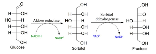
Glycine at a dose of approximately 10 g per day should block the activation of aldose reductase, if glucose levels rise because glucose is not being processed quickly enough due to a shortage of NAD+ molecules.
Flurbiprofen (Strepfen), in a small amount (less than the amount used for a sore throat; I estimate approximately 1 to 2 doses of 8.75 mg per day), may help activate the enzyme ALDH2, which breaks down the HNE aldehyde that has formed.
We should probably allow all these measures to act simultaneously to protect the liver and kidneys from damage during increased release of LA from adipose tissue. In the mouse model, we saw what this does. Unfortunately, we only have studies examining the individual effects of each method separately. It would be very interesting to see the results of blue-light exposure with these protective measures applied together, along with an analysis of the composition of stored fat.
Next
References:
Kinetics of the depletion of linoleic acid in mice
The Hidden Cost of Blue Light: Multiorgan Pathology Induced by LED Exposure in Mice


Comments
Post a Comment