Do omega-6 fats cause inflammation via aldose reductase and fructose?
So here I have another quite provocative headline. Don’t you think? The fact that omega-6 polyunsaturated fats, specifically linoleic acid contained in vegetable seed oils, promote the development of chronic inflammatory processes is generally well known. There is nothing provocative about that. But that this process could take place through the polyol pathway? Or even through endogenous fructose production? That, I think, is not generally known. Is it really so?
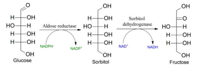
To demonstrate this, we must first accept that the linoleic acid peroxidation product 4-hydroxy-2-nonenal (HNE) is capable of activating the production of an enzyme called aldose reductase (AR). This enzyme is able to degrade HNE, preventing its further accumulation. If we suppress AR activity, for example with sorbinil, HNE accumulates, as a study has shown. The whole mechanism works as follows: if there is a reduction in antioxidant protection within the cell (for example due to fructose via the shutdown of SIRT2), the production and release of HNE increases, AR is activated, and oxidized residues of polyunsaturated oils are removed.
 |
| Suppression of aldose reductase activity (Sorbinil) causes an increase in HNE levels, thus blocking its removal. |
 |
| The aldehyde HNE, derived from linoleic acid, activates the expression of aldose reductase (AR), the entry enzyme of the polyol pathway that produces fructose from glucose. |
However, this has a number of side effects that are directly related to AR. It is a multifunctional enzyme, as we will see further. Pharmacological inhibition of the AR enzyme suppresses inflammatory markers, suppresses the response to endotoxins (LPS), and suppresses the activity of COX enzymes—essentially all signaling that is used in research as inflammation signaling. It therefore appears that the polyol pathway is involved in triggering inflammation not only in the vascular endothelium, but also, for example, in the retinal tissue of the eye, specifically in supporting cells of the nervous system, the so-called glial cells, or even in the cells covering the eye lens.


 |
| Suppression of AR activity using Sorbinil suppresses the induction of inflammation caused by high glucose levels or inflammation induced as a response to bacterial endotoxins (LPS), preventing the formation of inflammatory signaling molecules. |
What is interesting is that none of these studies mentioned fructose. We therefore do not know whether inhibition of fructokinase (KHK) would have the same effect. I therefore cannot claim that what they observed is an effect of fructose and activation of the KHK enzyme—unfortunately. I think that it is. That it is a general principle of regulating glucose levels within the cell.
Increased AR activity causes greater sensitivity to external stimuli—bacteria, endotoxins, glucose levels, and so on. So the key question is: what activates aldose reductase? Based on other research, I believe that in addition to high glucose levels, it is primarily omega-6 polyunsaturated fats released from membranes and peroxidized by oxidative stress, as I described in the vicious cycle of obesity. Activation of AR is probably the most important mechanism regulating inflammatory responses and their sensitivity to external stimuli. However, I do not recommend inhibiting AR; we have other methods to suppress inflammation.
References:
Acute Hyperglycemia-Induced Inflammation in MIO-M1 Cells: The Role of Aldose Reductase
ALDOSE REDUCTASE: New Insights for an Old Enzyme
Aldose reductase functions as a detoxification system for lipid peroxidation products in vasculitis


Comments
Post a Comment