Could a Deficiency of LA in Cardiolipin Be Caused by an Excess of LA?
Could an excess of the omega-6 fatty acid linoleic acid in the diet and in adipose tissue create the paradoxical situation in which linoleic acid (LA) is lacking in the phospholipids needed to form mitochondrial cardiolipin? That would mean reduced mitochondrial function and increased oxidative stress, which we observe in animal models fed very high-fat diets.
We have already encountered a similar situation with omega-3 fats. Take a look. The percentage of omega-3 phospholipids in cell membranes initially increases sharply but then declines as the consumption of more plant-derived omega-3 and omega-6 fats increases. This phenomenon is explained by changes in the activity of the enzymes that elongate and desaturate plant fats into the long-chain fatty acids EPA and DHA, which are found primarily in fish oil. The same enzymes also elongate and desaturate linoleic acid (LA) into arachidonic acid (ARA), whose concentration is maintained relatively stable. Thus, a higher intake of LA means lower enzyme activity and slower production of both omega-3 and omega-6 phospholipids. A higher intake of ALA behaves similarly. We can see this in the graph from a mouse study, but it also applies to humans. However, the previous article also introduced another mechanism.

In the case of cardiolipin, however, the issue is probably different. Phospholipid synthesis is likely not the determining factor here; rather, it is the rate at which free fatty acids are released from phospholipids. When linoleic acid is rapidly released during acute oxidative stress, it is apparently LA released from mitochondrial membrane phospholipids—specifically from cardiolipin, which is composed of up to 90% LA. This is therefore the primary source of the well-known aldehyde HNE, that is, peroxidized linoleic acid. The enzyme that releases this particular LA is called iPLA2γ.

A deficiency of LA, i.e. below 1.5%, will not provide enough LA for the production of cardiolipin. But an excess of LA, i.e. its burning into ATP energy, will increase the production of H2O2, i.e. trigger the activity of the enzyme iPLA2γ and thus deplete membrane phospholipids of LA. There is then not enough raw material for the production of cardiolipins or even LA is released from cardiolipins. Mitochondria then cannot function properly.

Only an optimal amount of LA in the diet (approximately 2% of daily energy intake) will ensure healthy mitochondria.

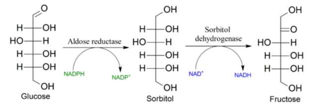
What happens if we genetically knock out iPLA2γ in a mouse model? When we preserve LA in cardiolipins. When we deprive mitochondria of the main source of HNE, an activator of the polyol pathway and a source of oxidative stress. You may find it surprising, but the mice become completely resistant to weight gain on a high-fat diet.

Perhaps even more surprising is how differently individual tissues respond to the knockout of iPLA2γ. Fat cells remain small and highly functional. They burn both fat and glucose very well and produce heat.

In contrast, muscle cells without iPLA2γ suffer from mitochondrial dysfunction. They are incapable of high performance and do not burn either fat or glucose well. Overall, the mice are smaller.
LA released from phospholipids allows for increased muscle performance, and a short-term increase in ROS can then act as a "boost".

A major difference is also seen in insulin production. Knocking out iPLA2γ reduces glucose-stimulated insulin secretion (GSIS). LA released from phospholipids strongly promotes insulin secretion, but when iPLA2γ is knocked out, LA is not released and insulin secretion is lower. This apparently results in resistance to weight gain and also protects fat cells from overload and poisoning by the aldehyde HNE, because HNE is no longer produced. This preserves their functionality.

This is simply very interesting. Knocking out iPLA2γ prevents cardiolipin repair in mitochondria. The primary function of iPLA2γ is to remove oxidized phospholipid acyl chains, allowing them to be replaced with fresh, intact LA chains. Peroxides activate iPLA2γ and thereby switch on their own repair mechanisms.
So if iPLA2γ is knocked out, muscles cannot renew peroxidized cardiolipin and therefore do not have enough energy. But why does this not affect fat cells? That remains quite a mystery. I can only speculate. The lower insulin level probably reduces oxidative stress enough to prevent cardiolipin from being damaged by peroxidation. It likely remains intact and does not degrade.
At the same time, we also see that all the enzymes involved in fat synthesis and fat storage are highly active in adipose tissue, yet this does not cause obesity. I have emphasized this here several times before: easy fat storage does not cause obesity. Difficult fat storage does.
The idea that consuming more linoleic acid will enrich cardiolipin stores is completely detached from reality. On the contrary, limiting linoleic acid intake to an optimal level and eliminating oxidative stress caused by aldehyde toxicity—that is, ensuring adequate aldehyde dehydrogenase (ALDH2) activity and possibly inhibiting aldose reductase (AR)—may help ensure that linoleic acid remains where it belongs, in cardiolipins, allowing efficient ATP production through the oxidative phosphorylation of the foods we eat.
Monitor the Rate of Processes—Fast Does Not Mean Safe!
References:


Comments
Post a Comment